
MEDICAL IMAGING INTERFACE
Diagnostic Visualization System
SCIENCE, TECHNOLOGY, VISION
Where Clinical Practice and Design Converge
SCIENCE, TECHNOLOGY, VISION
Where Clinical Practice and Design Converge
In diagnostic imaging, every visual detail contributes to the quality of the clinical assessment.
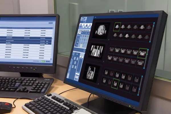
Elena Lazzara oversaw the Art Direction, Visual Strategy, and the entire UX/UI Design of the software interface for diagnostic imaging.
This was developed in collaboration with Zero Division, a studio specializing in artificial intelligence, computer vision, and blockchain technologies, and with the Department of Diagnostic Medicine and Radiology at the Policlinico Umberto I in Rome, a leading institution for diagnostic imaging and nuclear medicine.
PET, SPECT, AND MRI
From Plane to
the Third Dimension
PET, SPECT, AND MRI
From Plane to the Third Dimension
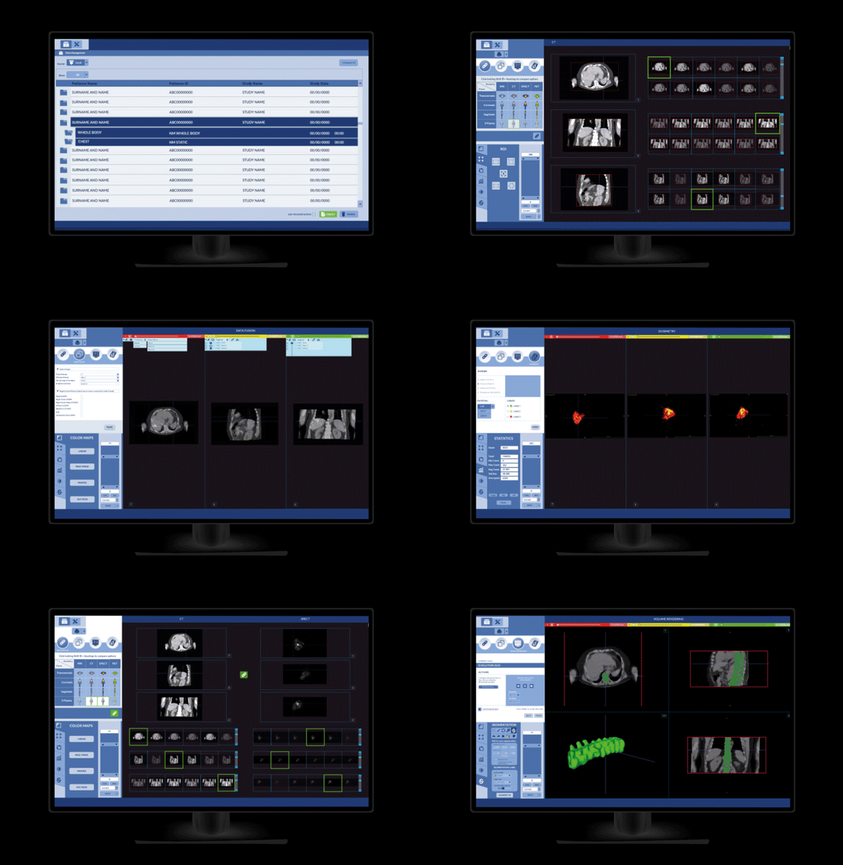
The project stems from a specific clinical need: to provide physicians with a tool capable of transforming complex PET, SPECT, and Magnetic Resonance Imaging acquisitions into a clear, consistent, and immediately interpretable visualization.
The system acquires images across the three anatomical planes — coronal, sagittal, and frontal — allowing the clinician to select the most significant captures, which the interface automatically reconstructs into a highly legible three-dimensional visualization.
MONITORING CLINICAL EVOLUTION
The Dimension of Diagnosis
A central feature of the system is the longitudinal comparison function: 3D reconstructions can be compared with datasets acquired at earlier times to precisely detect the patient’s clinical evolution, whether regarding changes, improvements, or stability in the diagnostic presentation.
A seemingly simple detail that actually requires an information architecture capable of supporting data complexity without ever sacrificing visual clarity.

MONITORING CLINICAL EVOLUTION
The Dimension of Diagnosis
A central feature of the system is the longitudinal comparison function: 3D reconstructions can be compared with datasets acquired at earlier times to precisely detect the patient’s clinical evolution, whether regarding changes, improvements, or stability in the diagnostic presentation.
A seemingly simple detail that actually requires an information architecture capable of supporting data complexity without ever sacrificing visual clarity.


ICONS, BUTTONS, INTERFACE
A Coherent Grammar
ICONS, BUTTONS, INTERFACE
A Coherent Grammar
Elena Lazzara’s work focused on the complete redefinition of the graphic system: the redesign of all functional icons, control buttons, master images relative to the three anatomical planes, and the reorganization of the entire multi-window interface.
Every choice — from the information hierarchy to the functional palette, from the iconographic language to the interaction patterns — was calibrated to reduce the physician’s cognitive load and accelerate the decision-making process during the diagnostic phase, without ever compromising clinical accuracy.



SPHERICAL METHOD IN ACTION
SPHERICAL METHOD
IN ACTION
In the Medical Imaging interface project, the Spherical Method was applied to transform a vast quantity of diagnostic data into a visual system capable of supporting clinical reasoning.
The approach connected medical imaging, information architecture, visual perception, cognitive ergonomics, and operational workflows, designing an interface in which every element contributes to the understanding of the diagnostic picture.
The three-dimensional reconstruction, navigation between the different anatomical planes, and the organization of information were not conceived as isolated features, but as parts of a single design ecosystem that helps physicians interpret complex images with greater speed, continuity, and precision.